muscle fiber orientation
5 Things to Remember 1. There are many possible ways to specify the complex fibre orientations in a finite element model for example defining a local element coordinate system.

Myocardial Mechanics Structure And Function Of Myocardial Fibers Ecg Echo
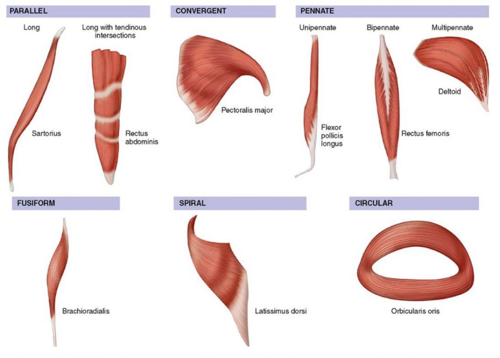
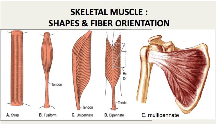
Tending to approach each other.

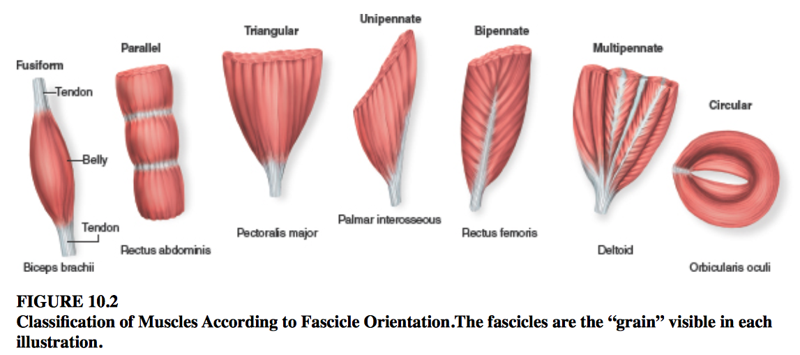
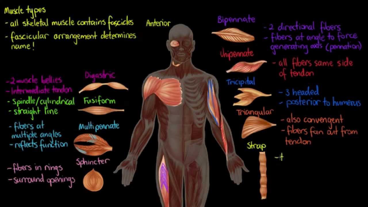
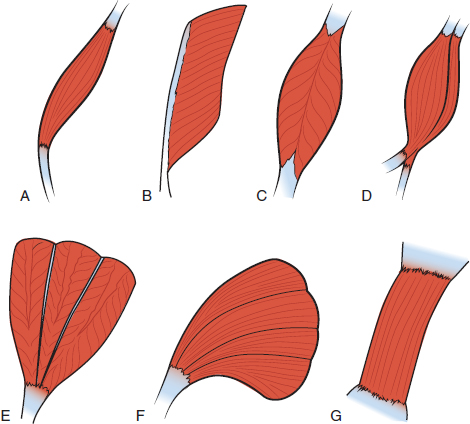
. Abstract Muscle fiber orientation in the left ventricular myocardial layer was histometrically estimated in normal concentric and eccentric hypertrophied hearts. The arrangement of a T-tubule with the membranes of SR on either side is called a triad Figure 1032. Unipennate muscles are those where the muscle fibers are oriented at one fiber angle to the force-generating axis and are all on the same side of a tendon.
This study was designed to locate the middle of the muscle fibers of commonly injected muscles thus identifying the endplate zone of these muscles. Comparisons between CFD and Diffusion Tensor Imaging. Actions associated with abdominal muscle control can be complex.
The automatic methods proposed in recent years also involved voting procedures which were computationally expensive. The angle of inclination of muscle fibers from coronal section was largest in the innermost and outermost zones and was progressively diminished toward the middle layer in all the hearts. The metabolite profiles were different for each orientation of muscle fibers to the main magnetic field.
Tending to one point of focus. The proximal and distal musculotendinous junctions in muscles of the upper and lower extremities were identified. In addition the complex fibre orientation arrangement makes it quite difficult to create an accurate finite element muscle model.
Muscle fiber orientation and connective tissue content in the hypertrophied human heart To elucidate the structural correlates of cardiac failure in myocardial tissue muscle fiber alignment and connective tissue volume fraction were measured at multiple sites in the left ventricular free wall and in the interventricular septum of 14 human hearts. Orientation of muscle fibers was determined. Some textbooks include Fusiform muscles in the parallel group.
Ward in Comprehensive Biomedical Physics 2014 101122 Recruitment and Summation. CFD Simulations for 3D Muscle Fiber Architecture in Finite Element Analysis. When the muscle fibers are stimulated indirectly via their nerve supply the muscle fibers are activated synchronously because their motoneurons are activated simultaneously.
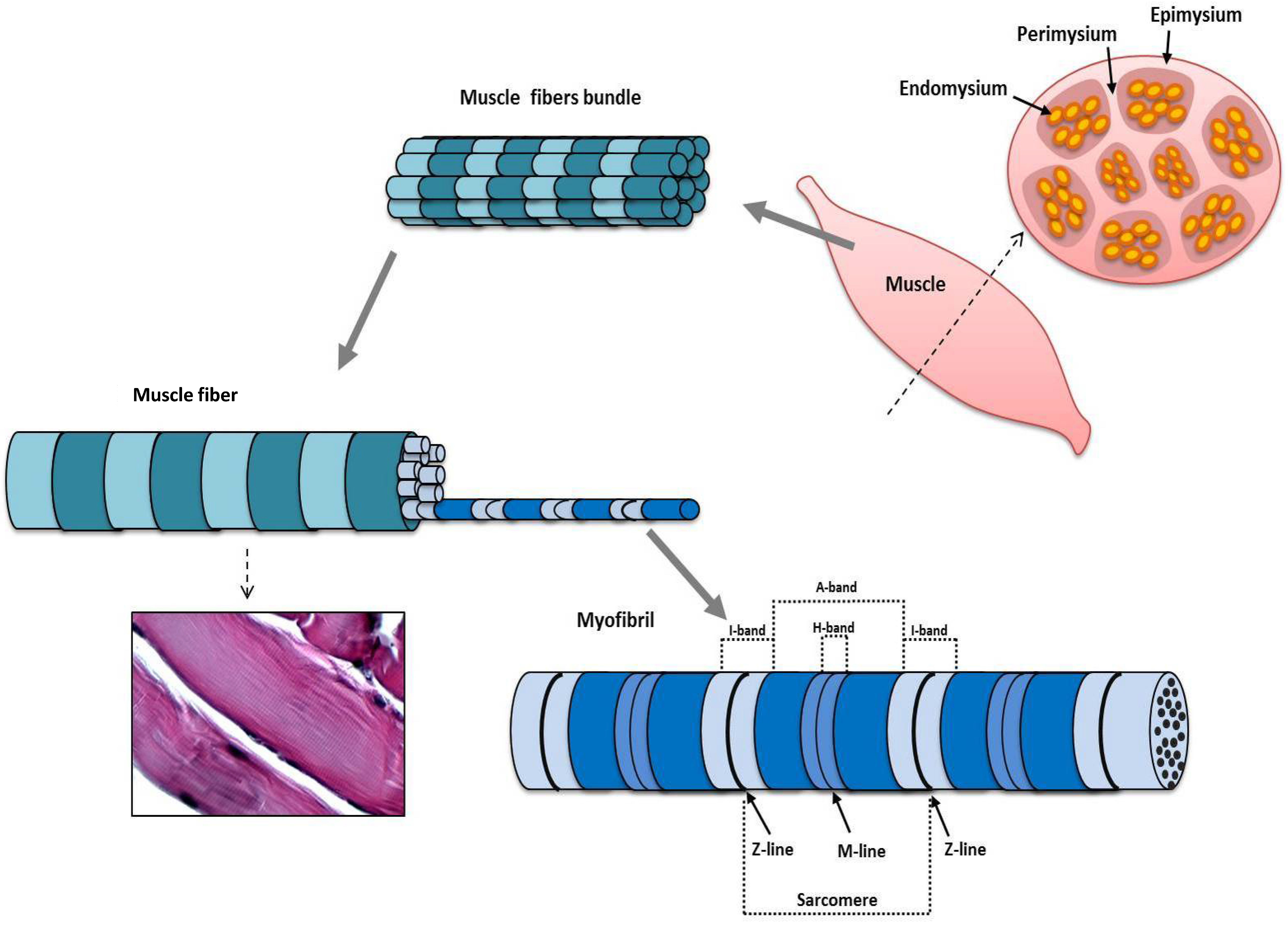
Because the diameter of a muscle fiber can be up to 100 μ m the T-tubules ensure that the action potential on the membrane can get to the interior of the cell and close to the SR throughout the sarcoplasm. The metabolite profile changes due to the muscle fiber orientation demonstrate that the positioning potentially causes inaccuracy in 1 H-MRS spectrum analysis. A single muscle does not usually work in isolation but in harmony with others.
This fiber orientation provides a good compromise in providing for both range. They are normally long muscles which cause large movements are not very strong but have good endurance. This study reveals that the muscle orientation at 0 30 60 and 90 to the main magnetic field significantly affects the metabolite profile and quantification.
Orientation of muscle fibers was determined. Muscle fibers are typically large cells some 20100 μm in diameter and many centimeters long with the longest fibers being about 12 cm. All the abdominal muscles have different muscle fibers orientation and act in all three planes during movements and are linked together by having a common site of connection or by fascia.
Orientation of muscle fibers was determined. The traditional manual method for MFO estimation in sonograms was labor-intensive. The orientation at 90 was the most different compared to other orientations.
Examples include Sartorius and Sternocleidomastoid. The pennation angle in unipennate muscles has been measured at a variety of resting length and typically varies from 0 to 30. Endurance training has minimal effect on the size of muscle however it does increase mitochondrial mass allowing for increased oxidative metabolism in skeletal muscle.
These dark lines are surrounded by connective tissues which appear bright. For optimal pick-up of electromyographic EMG signals surface electrodes are best aligned in parallel with the fibre orientation of the underlying muscle. The quantity of IMCL and EMCL exhibited statistically significantly changes with impacts at 30 60 and 90 when compared with muscles aligned at 0 to the main magnetic field.
Each stimulus pulse activates a proportion of the fibers in the nerve trunk and the activated fibers. Measurements using common surface landmarks were used to determine the relationship of these muscles with the landmarks eg biceps muscle bulk extends from the upper fourth to the lower fourth of the humerus. In the case of muscle fibers within an ultrasound image they appear as dark parallel lines with slight curvature at particular angles typically 530 within the muscle belly.
Knowledge of skeletal muscle fiber orientations is important for modeling mechanical properties and behavior of muscle tissue. Measurements using common surface landmarks were used to determine the relationship of these muscles with the landmarks eg biceps muscle bulk extends from the upper fourth to the lower fourth of the humerus. This study aimed to measure muscle fibre orientation and other parameters of muscle morphology of the abdominal muscles in relation to palpable bony landmarks.
At the level of ASIS the muscle fibres of obliquus internus abdominis were almost horizontally orientated but at 2 cm below ASIS were aligned about 6 degrees inferomedially to the horizontal. The dense muscle fiber microstructure gives rise to orientation dependent mr features with anisotropic overall motion of the creatine cr and phosphocreatine pcr molecules causing residual dipolar couplings first described for the total observed creatine tcr crpcr resonances 1 2 while orientation dependence was later also reported for. A skeletal muscle fiber arrangement in which the fibers are in an somewhat parallel orientation spread across a wide bone surface at the origin and coming together at a tendon attachment at the insertion.
Muscle fiber orientation STUDY PLAY parallel strap have fibres which as the name suggests run parallel to each other. There are three different muscle fiber typesslow oxidative fast oxidativeglycoltic and fast glycolytic. Skeletal muscle tissues have complex geometries.
Muscle fiber orientation MFO is an important parameter related to musculoskeletal functions. These cells are multinucleated because they need many nuclei to govern protein synthesis and degradation.

Skeletal Muscle Parts And Classification Fascicular Arrangement Anatomy Qa
Changing The Way You Learn Flashcards

Fascicle An Overview Sciencedirect Topics
Muscle Types Youtube
Jfmk Free Full Text Morphological And Functional Aspects Of Human Skeletal Muscle Html

Skeletal Muscle Shapes Fusiform Muscles Thick In Middle And Tapered At Ends Parallel Muscles Have Parallel Muscle Fibers Convergent Muscle Broad At Ppt Download

11 2 Explain The Organization Of Muscle Fascicles And Their Role In Generating Force Anatomy Physiology

Pennate Muscle An Overview Sciencedirect Topics
Imaging Muscle Radiology Key
How Muscles Work Part 2 Of 2 Shapelog
Muscle Histology Flashcards Chegg Com

Myocardial Fiber Orientation And Direction Of Rotation Myocardial Download Scientific Diagram

Pennate Muscle An Overview Sciencedirect Topics

Organization Of Skeletal Muscles Course Hero

Classification Of Muscles Based On The Directions Of Muscle Fibers The Download Scientific Diagram

3 Orientation Of Cardiac Muscle Fibers Download Scientific Diagram
2
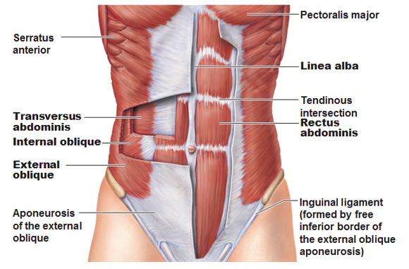
Muscles Of The Abdominal Wall
Organization Of Skeletal Muscles Course Hero
0 Response to "muscle fiber orientation"
Post a Comment